A large cavity does not automatically mean a child needs a pulpectomy. The deciding factors are the condition of the pulp, the tooth’s remaining structure, the roots, the surrounding bone and the developing permanent tooth underneath. By the end, you will know which symptoms require prompt assessment, how dentists compare a filling, pulpotomy, pulpectomy and extraction, and why follow-up matters.
Key takeaways
- Deep decay does not automatically mean a pulpectomy is needed.
- Seek prompt care for swelling, spontaneous pain, fever or a gum abscess.
- A healthy pulp may need a filling; infected pulp may need pulpectomy or extraction.
- Follow-up visits check healing and protect the developing permanent tooth.
What deep decay means and which symptoms need prompt attention
A cavity becomes concerning when decay passes through enamel and dentine toward the pulp, the inner tissue containing nerves and blood vessels. Deep tooth decay in children does not automatically require pulpectomy: a large cavity can still have a healthy or recoverable pulp.
The warning is a change in symptoms or signs suggesting irreversible inflammation, pulp infection, or tissue death.
| Feature | More consistent with enamel or dentine decay | More concerning for pulp involvement |
|---|---|---|
| Pain pattern | Brief sensitivity to cold or sweets that stops quickly | Spontaneous toothache, lingering pain after hot or cold food, or pain that wakes a child at night |
| Pressure | Little or no pain when biting | Pain on biting or tenderness when the tooth is tapped |
| Gum and face | No swelling or drainage | Gum swelling, a gum boil, pus, facial swelling, or fever |
| Tooth appearance | Visible cavity without colour change | A darkened tooth, especially with a large cavity |
| Symptoms | Discomfort linked to eating | No pain does not rule out infection; a draining gum boil can occur without toothache |
Arrange prompt dental examination for spontaneous toothache, pain that wakes a child at night, swelling, fever, drainage, or facial swelling. A dentist checks the tooth, gum, tenderness, and drainage, then uses a dental radiograph to examine the roots, surrounding bone, and developing permanent tooth. Symptoms alone cannot confirm the pulp’s condition.
How a paediatric dentist determines the condition of the pulp
Clinical examination starts by checking whether the tooth is restorable and whether tapping or biting causes tenderness. A pulp vitality test, when a child can respond reliably, helps interpret the tooth’s reaction, but no single result proves that the pulp is healthy or dead.
A dental radiograph for baby teeth adds essential evidence because symptoms alone cannot reliably show pulp status.
- Reversible pulpitis: brief pain from cold or sweets that stops quickly after the trigger is removed, without spontaneous pain, swelling, drainage, or root-area changes.
- Irreversible pulpitis: spontaneous pain, lingering pain after hot or cold food, night pain, or pain on biting. A pulp exposure that bleeds heavily or cannot be controlled during treatment also supports advanced inflammation.
- Dead or infected pulp: no response on testing, a gum boil, swelling, pus, tenderness, or a darkened tooth. Pain can disappear after the pulp dies.
The radiograph checks whether decay reaches the pulp and whether enough sound tooth remains for restoration. It also identifies furcation or periapical bone loss and internal or external root resorption. The image shows the developing permanent successor and helps assess its position.
A vital tooth with inflamed coronal pulp but healthy radicular pulp may fit pulpotomy. Necrosis, suppuration, or infection extending into the canals supports pulpectomy when the tooth remains restorable; severe destruction or an unmanageable infection can make extraction safer.
When a filling, pulpotomy, pulpectomy or extraction is appropriate
A filling is appropriate when decay has not infected the pulp and enough sound tooth remains for a sealed restoration. If the coronal pulp is inflamed but the root pulp remains healthy or reversibly inflamed, a pulpotomy removes only the affected upper portion.
Pulpotomy versus pulpectomy is therefore a question of how far the disease has spread.
| Option | What it preserves or removes | When it applies |
|---|---|---|
| Filling | Preserves the entire pulp; removes decay | The pulp is not irreversibly inflamed or infected, and the tooth is restorable |
| Pulpotomy | Removes the inflamed coronal pulp; preserves vital root pulp | The tooth remains vital, with no pus, necrosis, or root-canal infection |
| Pulpectomy | Removes pulp from the crown and root canals, then fills the canals | Pulpectomy indications include spontaneous or lingering pain, swelling, a sinus tract, abscess, furcation bone loss, necrosis, or pathologic root resorption |
| Extraction | Removes the primary tooth | The tooth cannot be predictably restored, has extensive root resorption or poor bone support, or is close to normal exfoliation |
A pulpectomy is not justified by cavity depth alone. The dentist combines symptoms, exposure findings, clinical examination, and radiographs before choosing it, and checks that the tooth can remain useful until the successor develops.
Primary tooth extraction criteria also include uncontrolled infection, inadequate remaining root structure, or a position that risks the developing permanent tooth. A child can have a dead or infected pulp without pain, so absence of symptoms does not automatically favour a filling or pulpotomy.
What primary-tooth pulpectomy involves and how it protects the successor tooth
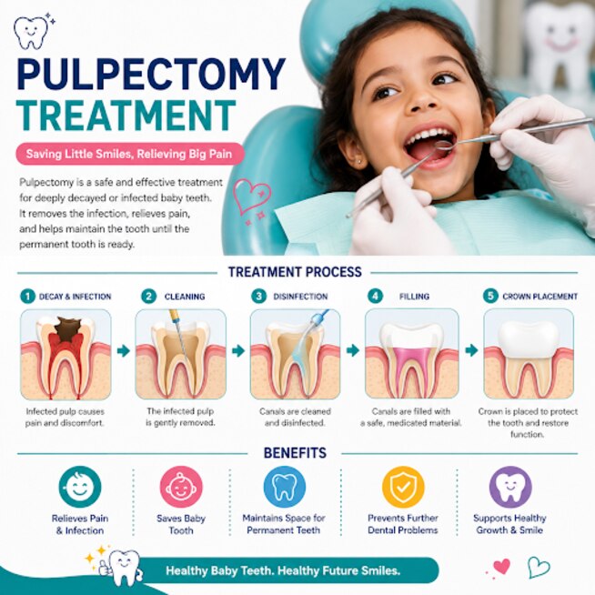
A pulpectomy removes infected or irreversibly damaged pulp from a restorable primary tooth. After opening the tooth, the dentist cleans and shapes each canal, measures its working length, and uses clinical findings and radiographs to avoid over-instrumentation. Cleaning must stop short of the developing permanent tooth and surrounding tissues.
Using a rubber dam for pulpectomy keeps saliva and bacteria away from the canals, improves visibility, and protects the child from instruments and irrigants. The canals are then filled with a resorbable root canal filling material designed to disappear as the primary tooth’s roots resorb.
Permanent-tooth gutta-percha is not automatically suitable: a material that remains while the successor erupts can interfere with normal development. Extruded filling material, uncontrolled instruments, or untreated infection can also harm the developing permanent tooth.
The tooth needs a durable restoration after the canals are sealed, often a full-coverage restoration when substantial structure has been lost. A weak or leaking filling lets bacteria re-enter and can turn a technically successful pulpectomy into another infection. Keeping the primary tooth can preserve chewing function, arch space, and guidance for its successor.
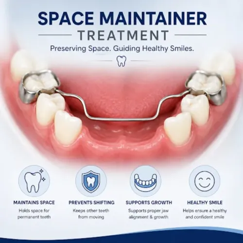
If the tooth must be extracted, ask whether a space maintainer after extraction is needed; the decision depends on the child’s age, tooth, successor development, and bite.
Why review visits matter after treatment
A treated primary tooth still needs pulpectomy follow-up because infection or root changes can develop without pain. At home, watch for:
- Renewed toothache or pain on biting
- A gum boil, draining sinus, or recurrent swelling after pulpectomy
- Facial swelling, fever, bad taste, or increasing looseness
- A filling or crown that breaks, feels high, or stops the child chewing comfortably
Arrange reassessment promptly if any of these signs appear. Do not wait for the next appointment if swelling spreads, fever develops, or your child seems unwell; antibiotics alone do not remove infected canal tissue, while spreading infection may require antibiotics alongside urgent dental treatment.
The dentist should examine the tooth and take a follow-up radiograph when indicated, commonly at a six-month dental review and periodically until the tooth exfoliates. These checks look for healing, new furcation or periapical radiolucency, abnormal root resorption, mobility, and effects on the developing successor tooth.
A paediatric dentist in Hadapsar can compare symptoms, restoration, radiographs, root development, and expected exfoliation before choosing observation, retreatment, or extraction. Dr Rohini Lahane can provide local continuity, helping parents distinguish a change needing prompt advice from one suitable for scheduled follow-up.
Related service
Pediatric / Kids Dental Care by Dr. Rohini Lahane in Hadapsar, Pune Dr. Rohini Lahane (BDS, MDS) offers gentle and effective Pulpectomy Treatment for children to save severely... View service → |
Frequently asked questions
What symptoms from deep tooth decay need prompt attention?
Seek prompt dental care for spontaneous or night pain, sensitivity that lingers, facial swelling, a gum abscess, fever, difficulty eating or reduced mouth opening.
How does a paediatric dentist assess the pulp?
The dentist examines the cavity, asks about pain, checks sensitivity and tenderness, and reviews dental X-rays for infection, bone changes and the condition of the developing successor tooth.
When is a filling, pulpotomy, pulpectomy or extraction used?
A filling suits a tooth with a healthy pulp. A pulpotomy removes inflamed pulp from the crown when the roots remain suitable. A pulpectomy removes infected or irreversibly inflamed pulp from the crown and roots. Extraction is considered when the tooth cannot be restored, infection cannot be controlled or too little tooth remains.
What does a primary-tooth pulpectomy involve?
The dentist removes infected pulp from the baby tooth’s crown and roots, cleans and fills the canals with a resorbable material, then seals the tooth with a restoration. Removing infection helps protect the developing permanent tooth.
Why are review visits important after pulpectomy treatment?
Reviews check the restoration, pain, swelling and gum tissues, and dental X-rays can monitor healing and the developing permanent tooth until the baby tooth naturally sheds.